RSS

Category Archives: Blood Analysis

Q and A: How Would My 1925 Detective Determine That a Stain Was Human Blood?

Q: The setting is rural 1925. There are dark stains on trees, shrubs and leaves which my hero believes is blood. My questions are, how would he identify it as blood and how would he discriminate it from animal blood? What tests or experiments that existed in that era could he perform?

Frank James, Ste-Marthe-sur-le-Lac, Canada

A: The two steps needed to distinguish animal blood from human blood are: Determining if the stain or sample is indeed blood and then is it human of animal.

Testing liquids and stains to determine if they are blood is not new. For centuries, the microscope has been used to visually identify blood cells, which would prove that the substance is blood. But this required liquid blood and not the typical crime scene clotted or dried blood, neither of which contain identifiable cells. Several other tests appeared in the 1800s, including the hematin test, developed by Polish scientist Ludwig Teichmann in 1853. This also required liquid blood since in this test the blood was mixed with acetic acid and salt crystals, heated, and then viewed under a microscope. The presence of the characteristic rhomboid crystals proved the sample was blood. This test is similar to the present day Teichmann and Takayama Tests.

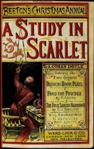
The guaiacum test, developed in 1862 by Dutch scientist Izaak Van Deen, used the guaiac resin of a West Indian shrub and is the precursor of the present day phenolphthalein test (see below). In the guaiacum test, the blood sample was mixed with hydrogen peroxide and guaiacum and, if it was indeed blood, a blue color would appear. In 1887, a similar test was used by Sherlock Holmes to identify a bloodstain in the very first Holmes story, A Study in Scarlet.


In 1900, Paul Uhlenhuth developed a serum that reacted only to human blood, and not animal blood. This is an antigen-antibody reaction and is similar to how this testing is done today. The sample would be dissolved in salt water and then the serum would be added. Human blood proteins would then react with the serum, producing complexes that would precipitate (fallout of solution) and darken the serum. Animal blood would cause no such reaction so if a reaction occurred the tester would know that the blood was indeed human and if not it must be animal blood or some other substance. Now we have serums that react with a just about any species of animal you can name and with these lab techs can determine the specific type of animal that shed the blood.

So your character could use guaiacum to determine that the sample was blood and then employ Uhlenhuth’s serum to determine if it was human or not.


Q and A: What Common Substance Could My Amateur Sleuth Use to Determine If a Stain is Blood?

Q: In my next mystery an amateur sleuth finds a stain on a wood floor under a rug. Is there a common substance she could use to determine if it were blood?

SCurran, Monroe, WI

A: There are a couple of easily obtainable chemicals that could be used for this. The first is phenolphthalein which was one of the first chemicals used to test for the presence of blood. It is readily available in most pharmacies and can be ordered online. There is a link below so you can see a company that sells the stuff and what it looks like.


Another thing that is purchased in any pharmacy are urine testing strips. These test for many chemicals but one of the things they test for the blood. There are several absorbent squares on the test strip one of which is a test for blood. Simply moistened the strip or moistened the staying and press the strip against the reaction will occur very quickly and this is a sensitive test for even small amounts of blood. Definitive testing to determine if it is indeed blood or if it is human blood or love a more sophisticated but these two testing methods are highly sensitive for even very small amounts of blood.

 
Leave a comment

Posted by on December 5, 2010 in Blood Analysis


Blood Camera: The New Luminol?

The most important material that investigators can find a crime scene is blood. It tells them a great deal. Blood is both a biological and a physical material. That is it has biological properties but it also behaves as a liquid. Employing both of these characteristics can be of great benefit to investigators.

Blood typing and DNA are usually readily done on crime scene samples unless they are so degraded that this type of testing is not possible. That is not often the case. Blood type and DNA can exclude some people and point the finger directly at others.

Blood behaving as a liquid can reveal to investigators how the crime occurred. Did the blood simply leak from a wound or did it spray from an arterial injury? Was it spattered as the result of a gunshot or blows to the head from a baseball bat? Did the victim standstill, lie on the floor, or walk or run away after the injury? The blood splatter pattern can reveal what happened and often where the various players in the crime were at the time the blood was shed. This can support or refute suspect and witness statements.

Often a killer will attempt to clean up a crime scene and scrub away the victim’s blood hoping that there will be no evidence of the crime remaining. Most of you are familiar with Luminol which has been employed in such cases. When properly used it can reveal blood splatter patterns even after the scene has been cleaned and indeed will often show the swipes and scrubs left behind by the cleaning utensil. It is highly sensitive and will find blood in the parts per billion.

Using Luminol requires that the area be sprayed with Luminol solution and then the room must be darkened and viewed using UV light. This can sometimes be cumbersome, particularly when attempting to evaluate a scene during the daytime. The windows in the room must be covered and all sources of light must be blocked out to get the best effect.

Add to this the fact that many other substances such as bleach, coffee, and rust can interfere with Luminol testing. Luminol can also damage the blood so that DNA testing will be less accurate.

Now it seems that a camera has been developed that will do the work of Luminol. It apparently uses pulses of infrared light and then measures the light reflected back to the camera. It filters out unwanted light wavelengths and concentrates on those consistent with blood proteins.

This is a very clever tool and hopefully it will work out to be as useful as it seems to be.

 
5 Comments

Posted by on November 29, 2010 in Blood Analysis, High Tech Forensics


Estimating Age From Crime Scene Blood

The police are called to a suspected crime scene, one where a murder has likely taken place. There is no body. There is no suspect. There are no witnesses. But a large blood stain is found at the scene. Whose blood is it?


Any police investigator will tell you that identifying victim is one of the first and most important steps in identifying the perpetrator. The simple reason is that most murders are committed by someone with some relationship to victim. A spouse, a friend, a coworker. But without a corpse, how can the victim be identified?

Since in this circumstance there would be no description of victim, the police would not know where to look. They would have no age, sex, size and weight, height, or any of the other physical details that might narrow their search for who the victim might be. Each one of these factors can help narrow the possibilities.

But what if they could determine that the victim was a teenager or a middle-aged male or an elderly female? DNA obtained from the blood could easily determine the sex but not the age of the victim. Until now. There appears to be a new test that just might reveal age from a crime scene blood sample. And least in broad terms.

A recent report in the journal Current Biology submitted by researchers from the Erasmus MC University Medical Center in Rotterdam, Netherlands suggests that byproducts from human T cells might supply this information. It’s complex biology but it seems that our T cells, which are an important component of our immune system, have great diversity in their receptors. It is these receptors that allow them to recognize a multitude of foreign invaders and tag them for destruction by the white blood cells and other components in the complex system that protects us from infections.

It seems that this diversity is accomplished through a constant rearrangement of the DNA within the T cells. A byproduct of this process is the creation of small circular DNA molecules known as Signal Joint TCR Excision Circles, or sjTRECs for short. It appears that the amount of these DNA packets declines at a constant rate with age. Using them, these researchers believe that they can narrow the age range of the person who shed the blood to within 20 years. Not very accurate but it would distinguish a teenager from a middle-aged person or an elderly individual and this in turn might help identify the victim.

Stay tuned. This could prove to be an interesting and useful technique. Or not.


Erythropoietin and Survival Time

In victims of traumatic deaths, one of the questions that is often useful to investigators is how long the victim lived after the traumatic event. Let’s say someone is in an automobile accident or is shot or stabbed and bleeds to death. Did this take 15 minutes or 15 hours? A hormone within the blood might help with this determination.

Erythropoietin (EPO), a hormone produced in the kidneys and liver, regulates red blood cell (RBC) production. In people who are anemic, that is who have a low RBC count, erythropoietin production is revved up so that more RBCs will be produced by the bone marrow. The body has a way of taking care of itself. Erythropoietin is also a performance-enhancing drug in that it increases red blood cell production and therefore increases the capacity of the blood to carry oxygen. The more oxygen the blood carries the longer and more intensely someone can exercise. Distance runners and cyclists have often used erythropoietin to improve their performance in races. It is banned by virtually every competitive organization in the world.

So what does all this have to do with survival time after an injury? It is been found that when someone is bleeding and their blood count is dropping because of this blood loss, the kidneys and the liver began to produce larger amounts of erythropoietin. All they see is that the red blood count is dropping and that the blood pressure is low and their natural response is to increase the production of this hormone.

A group of researchers at the Osaka City University Medical School’s Department of Legal Medicine recently published an article in the journal Forensic Science International in which they looked at the blood levels of EPO in relationship to survival time after major injuries that caused massive bleeding. They found that victims who survived many hours after such injuries showed a rapid increase in EPO in their blood serum over the first six hours after the injury. They concluded that this test might be useful in determining if someone died early within the six-hour window versus dying later.

How would this be useful in a criminal case? Remember the case of Chante Mallard? This 27-year-old woman in Fort Worth, Texas decided to get drunk and stoned and then drive home at around three o’clock in the morning. Unfortunately Mr. Gregory Biggs was walking along the road. She struck him with her car. He flew through the passenger side windshield and was lodged in the shattered window head down in her passenger seat. Ms. Mallard did what anyone would do in that circumstance: she drove home, parked the car in the garage, and did some more drugs, leaving Mr. Biggs to bleed to death.

Mr. Biggs survived the initial impact and only died later because Ms. Mallard failed to do the right thing and call for the medical care that might have saved his life. The evidence for this was that his blood pooled within the door pocket of Ms. Mallard’s car where his hand had come to rest. The fact that blood ran down his arm and collected in this area proved that he was still alive for many hours after the impact. At death, the heart stops, the blood ceases its movement, and bleeding stops.

Had this new EPO test been available and used in this case, it would’ve shown a very elevated level in Mr. Biggs whereas had he died almost immediately from the impact these levels would be very low. In the latter case, Ms. Mallard would be guilty of reckless homicide but the fact that she ignored this man and allowed him to die a slow death added another level of depravity to this situation. This might explain why she was sentenced to 50 years in prison rather than some shorter time.


Blood Shouts: Review of Blood Secrets by Katherine Ramsland

On June 12, 1994, a barking dog in the exclusive Brentwood enclave of Los Angeles alerted a neighbor to a scene that would soon garner headlines around the world: the double homicide of a young waiter, Ronald Goldman, and Nicole Brown Simpson, former wife of football great O. J. Simpson. Police went to Simpson’s home to check on his welfare and noted a bloodstain on the door of his white Ford Bronco. A trail of blood led up to the house, but Simpson had just flown to Chicago. When questioned, he denied having anything to do with it, although a fresh cut on his hand proved suspicious. Then several droplets of blood at the scene failed to show a match with Brown or Goldman. Their killer had cut himself.

Simpson’s blood was drawn for testing, which indicated that the unknown blood had three factors in common with Simpson’s and that only one person in 57 billion could produce an equivalent match. In addition, the blood was found near footprints made by a rare and expensive type of shoe—O. J.’s size. Next to the bodies was a bloodstained black leather glove that bore traces of fiber from Goldman’s jeans, and it matched a bloody glove found that night on his property. Traces of blood from both victims were lifted from it, as well as from inside Simpson’s car and house, along with blood that contained his DNA. His blood and Goldman’s were found mixed together on the car’s console. Simpson was arrested and charged.

Forensic serologists at the California Department of Justice, along with a private contractor, did the sophisticated DNA testing. Three crime labs determined that the DNA in the drops of blood at the scene matched Simpson’s.

Nevertheless, at Simpson’s trial the following year, criminalist Dr. Henry Lee testified that there appeared to be something wrong with the way the blood was packaged, leading the defense to propose that samples had been switched, blood had been planted, and the improper storage had degraded the samples past the point of accuracy.

The jury acquitted Simpson, and over a dozen books came out during the late 1990s from both sides to analyze the case.

Now, Rod Englert, a 46-year veteran of law enforcement, a homicide investigator, and an expert in blood spatter pattern analysis, has published Blood Secrets (St. Martin’s Press, April 2010; $25.99, co-wrtten with Kathy Passero). Among the many case evaluations he includes is the one he performed for the O. J. Simpson investigation. When assistant DA Marcia Clark invited his opinion, she told him “The crime is a goldmine for blood spatter analysis.”

Englert inspected every aspect of the crime and every significant surface and material, making fifteen separate trips to LA. He noted that almost all of the smears and spatters at the scene were sixteen inches from the ground or lower, which told him that the victims were on the ground when most of the bloodshed occurred. He surmised that Brown had been knocked unconscious and thus had not struggled with her attacker. The lack of blood on the bottom of her bare feet confirmed this. Goldman, on the other hand, had put up an enormous fight, fending off an aggressive knife attack. Because the back of his shirt was ripped but there were no wounds on his back, the killer “had wrenched Goldman’s shirt around almost backward in his effort to hang on to his victim.”

With blood evidence and information about the victims’ positions, Englert brainstormed with the team and reconstructed the scenario as this: “The killer had moved back and forth between his victims after they were incapacitated,” probably to ensure they were both dead. Before he fled, he cut Brown’s throat and punctured Goldman’s abdomen. It was also clear to Englert, after he ran several experiments with dogs, that Brown’s agitated Akita had probably walked through the blood, pawed at her, and possibly brushed against her in a protective stance.

Although Clark had expected Englert to testify, he took the stand. In this book, he provides the account he would have told the courtroom, as well as a quick assessment of what should the jury should have learned.

Just how Englert became a blood spatter analyst is, in itself, an unusual tale. To get readers there, he first describes his experience as a rookie cop, which led to his interest in learning how crime scenes are reconstructed.  Owning a cattle business on the side, he had a ready-made lab, as long as one could think outside the box…er, stall. He had cow’s blood, as much as he wanted, and a large barn to spatter it in. This chapter is well worth the read for any forensic scientist, if only to admire Englert’s innovations. “I dribbled blood from my fingertips,” he writes, “from the points of knives, and from holes in plastic garbage bags dragged across the barn floor. I tried the same tests on cement, gravel, dirt, sand, grass, wood and carpet to find out how the trails of blood differed….I made notes about how the blood got absorbed or distorted…” He shot into the blood with different types of guns, hit puddles of blood with bats, hammers, and boards, and scrutinized fine mist, thick drops, and cast-off patterns. He also wore different types of clothing to see how blood soaked into various materials.

Interpreting blood spatter patterns is a both a science and an art, but it can’t be fully learned from books. It requires practical hands-on experience and plenty of it. The shape of a blood drop can reveal a lot about the conditions in which it flew and fell, and Englert lays out the peculiar physics of blood spatter. When force is applied, for example, the amount of blood, shape of the drops, angle of impact, and location of a spatter at the crime scene will indicate everything from its velocity to the type of weapon used to how many people were involved. Blood with more weight travels farther, and it only travels so far in a straight line before it curves downward.

Bloodstain patterns help the investigators understand the positions and the means by which a victim and suspect moved, interacted, and possibly struggled through the scene. Investigators can then look for fingerprints, footprints, hairs, fibers and other forms of trace evidence. “I work my way backward through the chapters,” Englert writes “who, what, when, where, and how—until at last I reach the first page and find out how the story began.” In addition, an accurate reconstruction helps investigators determine which of witness and/or suspect is telling the truth.

Back to the Simpson case. Englert was certain that the blood evidence had provided “incontrovertible proof that Simpson had murdered Nicole Brown and Ronald Goldman.” He offers details, one item at a time, to back up his statement, focusing on Simpson’s socks, Goldman’s shoes, and Simpson’s car. He insists that despite claims of blood being planted, no one could plant that much blood spatter authentically enough to fool an experienced analyst. Englert fully demonstrates why this is so. “Truth,” he says, “got lost in the circus.”

In this engaging and readable book, Englert includes many different types of cases, some involving celebrities, some with a vexing mystery, and some from long ago, including a bullet trajectory analysis from the 1863 battle at Gettysburg.  He even admits to his errors, but the point of this book is to lay out the general process of blood spatter pattern analysis and show how each case has its own individual twists.

Ann Rule wrote the foreword and it’s clear that she’s familiar with Englert’s approach. She rightly says that “most of us involved in the circle of forensic science experts know one another…we are a motley crew, a fraternity who studies the blackest side of human nature and manages to find justice for victims of crimes and the truth for their survivors.” In fact, Englert has been involved in some of the cases about which Rule has written, and she had encouraged him to write a book. It’s no wonder that she’s pleased with the result.

There aren’t many casebooks available within the specific framework of blood spatter analysis; most are textbooks. Thus, Blood Secrets is different. It’s a full story about the life of a man who became an expert in one of crime’s most complex forms of evidence, and his analysis on many of the cases he’s worked. Thus, he gives readers a human side as much as an educational source – there’s something for true crime readers as well as for experts in the field. Blood spatter forensics has become an essential part of crime analysis, and the blood of victims will speak volumes about what happened to them when they can no longer speak for themselves.

Dr. Katherine Ramsland chairs the Department of Social Sciences at DeSales University in Pennsylvania, where she teaches forensic psychology and graduate-level criminal justice.

 
3 Comments

Posted by on April 13, 2010 in Blood Analysis, Guest Blogger


Mast Cells and the Timing of Wounds

An interesting article will appear in the October issue of the journal of Forensic Science International. It is titled: Mast cell reactivity at the margin of human skin wounds: An early cell marker of wound survival?

What?

Let me explain.

First of all, this article deals with very sophisticated biochemistry in that they measured the activity of certain esterase enzymes in relation to time after injury. I won’t attempt to explain it, but simply point out that research is ongoing in this distinctly forensic arena. But it does bring up the question of how the ME can judge whether a wound occurred pre-mortem or post-mortem.

This is sometimes one of the most difficult determinations the ME must make and his opinion impacts many things. For example, let’s say two assailants attack a victim. One shoots him three times in the abdomen, the other stabs him in the chest after he falls to the ground. Which injury killed him?

Sometimes it’s obvious from the nature of the wounds and the organs and blood vessels injured by each wound. At other times it’s not so clear. One key determination is whether the victim was dead before the stab wound occurred or not. If so, the second assailant might dodge a murder charge; if not, then the stab would could be the proximate cause of death or could at least be a contributing factor, in which case he could face serious charges.

The ME would analyze the stab wound. If it is clean and without irregular edges he can state that the victim was not wiggling or struggling at the time of the injury and just might have been dead beforehand. Living folks tend to fight back and try to avoid stab wounds and this struggle is often reflected in the character of the wound. Dead people tend to lie still so that the wounds are clean, in and out.

If there is no bruising or bleeding into or from the wound, the ME would know that the heart had stopped and there was no blood flow before the wound occurred. If bleeding occurred, the heart must have been beating and the blood flowing at the time of the injury.

Sometimes this is all he needs, but sometimes this type of evidence isn’t clear.

So, what are mast cells and what do they have to do with this?

Whenever an injury occurs, several types of cells rush the area to begin the healing process. Platelets in the blood arrive and begin to clump together and plug any breech in the blood vessels. This action is the first step in what we call the Clotting Cascade, and without it blood clotting would not occur and even minor injuries could cause lethal bleeding. White blood cells, lymphocytes, and mast cells also appear.

Mast cells contain little packets of chemicals–histamine and certain enzymes–in their cytoplasm–the liquid part of the cell. When an injury occurs the mast cells move in and release these chemicals. When properly stained and viewed under a microscope the cytoplasm of a mast cell is filled with tiny blue granules. These are the chemical packets. As the chemicals are released, the granules disappear, a process known as degranulation. This only happens while we are alive. After death the mast cells do not degranulate.

Mast Cells

Mast Cells: The large blue blobs are the nuclei and the tiny blue dots in the cytoplasm of these cells are the granules.

To determine if a wound was pre- or post-mortem, the ME can excise some tissue from near the wound edges, stain it, and look at it under the microscope. If the mast cells have degranulated he will know that the victim was alive at the time of the injury; if not, he will know just the opposite.

This type of examination is rarely needed, rarely done, and can offer confusing results in some cases, but it is an interesting technique for determining the timing of a victim’s wounds.

 
7 Comments

Posted by on September 11, 2009 in Blood Analysis, Time of Death, Trauma


5 Wild Recent Discoveries in Forensics by Rose Jensen

There are many things in science that seem more like science fiction and the study of forensics is no different. As soon as new methods of detection are developed, someone figures out a way to circumvent them, and new studies are constantly exposing new ways that criminals can be tracked, identified and caught. Here are some recent news stories on Forensics that expose just how interesting and dynamic it really can be and perhaps could provide some great fodder for those writing crime stories.

1–Detergents that wash away traces of blood. For the longest time, blood has been one of those substances that criminals could never quite get off their hands, or anything else, for that matter. With a little luminol or phenophthalene even the smallest traces could be brought to light. New detergents containing active oxygen are thought to work more effectively however, and actually remove all traces of blood making it much harder for forensic investigators to do their jobs.

2–Eating processed foods makes fingerprints more distinct. Crime fighters better hope their suspects have a penchant for fast food and fatty snacks. A forensic scientist at the University of Leicester has found that fingerprints with a higher salt content will leave more of a corrosive impression when put on metal. The higher the content of salt in a persons diet (a factor usually associated with heavily preserved foods) the easier his or her fingerprints are to see.

3–Bacteria have genomes too. While this might seem obvious, scientists are becoming better and better able to map and track these microbes many of which people carry naturally on their bodies and some of which can make people quite sick. Because these bacteria differ in their genetic makeup, some forensic scientists believe that they could potentially be the latest microscopic evidence to link criminals to their crimes.

4–Pollen could be the smoking gun. Pollen has long been studied by forensics experts, archaeologists and scientists as it is a microscopic but quite durable substance. A new use has been found for it, however, and scientists are tracking gun casings from the crime scene to suspects using a chemical cocktail. This mixture of chemicals varies uniquely depending on the weapon and coats any nearby pollen, creating a new link between the weapon and the shooter.

5–DNA testing just got a little faster. DNA testing has helped to solve numerous crimes and exonerate many innocent people, but it has long been an arduous waiting game because results can take some time to come by. Scientists in Japan are reporting that they’ve found a better way to do it that’s not only faster but more cost-effective as well, amplifying trace amounts of DNA into quantities that can easily be identified and linked to a suspect a process that has thus far been time consuming and costly.

This post was contributed by Rose Jensen, who writes about the associates degree. She welcomes your feedback at Rose.Jensen28@ yahoo.com

 
3 Comments

Posted by on September 2, 2009 in Blood Analysis, DNA, Guest Blogger